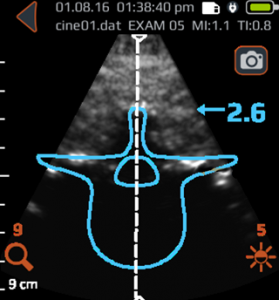
Notice the blue overlay on the Accuro display image; this indicates the spinous process cross-section is detected. When scanning a particular intervertebral level, if no orange interlaminar-space overlay appears, the interlaminar space will not be easily accessible with a midline approach.
Consider the following Accuro overlay indications:
Continuous blue overlay, despite angle and level, indicates an obstructed midline.
Accuro overlays show both spinous process and interlaminar space. (The ultrasound beam has a thickness, which means that it is possible to see both the spinous process and interlaminar space in the same image.)
In the above instances, identification of the interlaminar space may be attempted again after repositioning the patient or moving to a different intervertebral level.
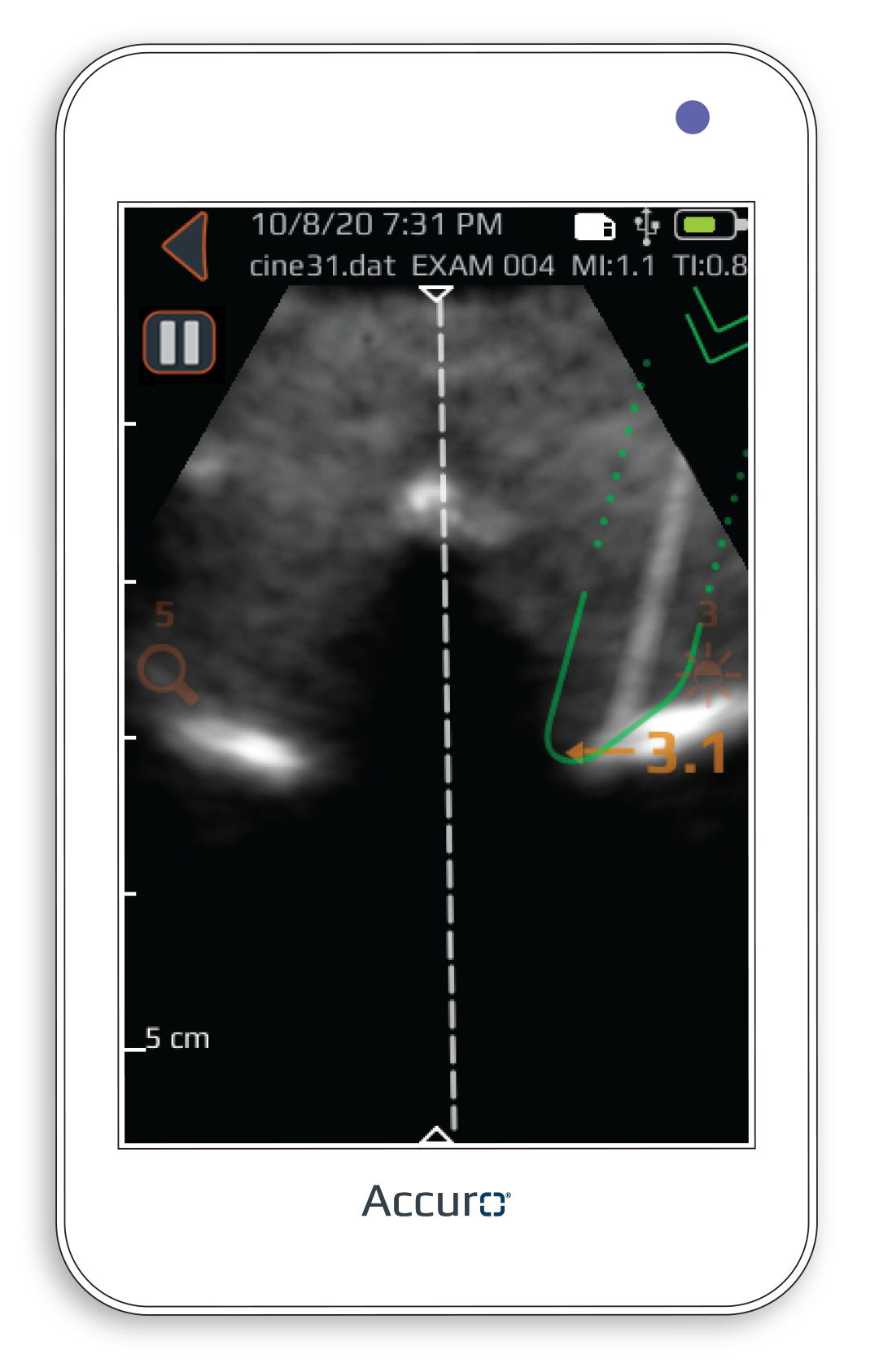
Alternatively, a paramedian approach may be attempted using the thoracic presetting, which identifies a prescribed paramedian needle path based on the Accuro Locator needle guide.
Accuro Display: Spinous Process
Thoracic Preset
Reasons for an obstructed view. Ligament calcifications appear as bone under the ultrasound, consequently obstructing the interlaminar view. Inaccessibility to the interlaminar space can be caused by spinal stenosis, osseous growths, or calcifications of surrounding ligaments. It occurs most often in the elderly population.
Obstructed interlaminar view
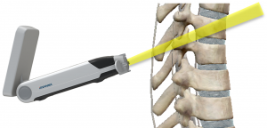
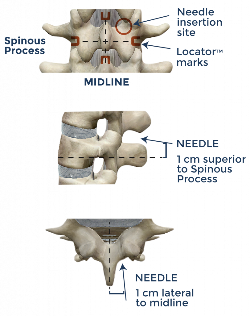
Paramedian lumbar epidural technique.
Accuro can be used to identify the spinous process position as a means to guide a paramedian lumbar epidural technique* as shown.
*Paramedian lumbar epidural protocol details from Ghosh et al., BJA Education, 16(7): 213-220 (2016)
Our website uses cookies so that we can provide you with the best user experience possible. Cookie information is stored in your browser and performs functions such as recognising you when you return to our website and helping our team to understand which sections of the website you find most interesting and useful. Thank you!
Strictly Necessary Cookies
Strictly Necessary Cookie should be enabled at all times so that we can save your preferences for cookie settings.
If you disable this cookie, we will not be able to save your preferences. This means that every time you visit this website you will need to enable or disable cookies again.

