
This content was reviewed by Stephen Garber, MD, Anesthesiologist in Southern California and updated in 2025 to reflect current clinical evidence and recent guidelines.
Anesthesiology is in flux. Particularly in the United States, demographics, economics, and technology are driving changes to care delivery mechanisms from the micro to the macro scale. The practice of anesthesiology is itself facing interesting trends — such as fewer resources and patients with greater need.
Conversely, the integration of advanced technology into everyday clinical practice offers real hope. The power of artificial intelligence (AI), for example, will redefine patient care standards. Central to this paradigm shift is RIVANNA’s mission to elevate global standards of care through the commercialization of world-first imaging-based medical technologies.
Below is a forecast of what’s to come in the future of neuraxial anesthesia — along with suggestions for influencing outcomes as this discipline and the roles surrounding it evolve rapidly.
Advancements in imaging and technology
Investigating technology plays an increasingly important role in elevating patient satisfaction and outcomes. The era of landmark-guided neuraxial anesthesia, relying solely on surface anatomy and palpation, could be ending. Imaging is no longer a supplemental option; it is becoming a core competency for clinicians performing epidurals and spinals. This shift is driven by both clinical needs and technological maturation.
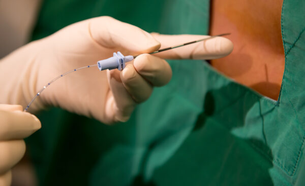
Ultrasound-guided techniques directly address these challenges. Real-time imaging techniques allow clinicians to visualize anatomical structures, like the epidural space, and infer the location of the ligamentum flavum and dura indirectly rather than relying solely on surface landmarks. In complex cases, ultrasound-guided placement consistently outperforms blind palpation in terms of first-pass success rates and needle placement times, all while reducing potential complications in neuraxial anesthesia.
The introduction of ultrasound guidance has overcome some of these barriers in important ways:
- Real-time imaging enables anesthesiologists to identify and target specific anatomical landmarks accurately.
- Complications and patient discomfort may be minimized through precise needle placement.
- First-attempt success rates are significantly higher with ultrasound guidance.
Importantly, device design has evolved alongside technique. Ultrasound systems intended for neuraxial applications are smaller, more intuitive, and designed for real-time procedural use at the bedside. Instead of repurposing large diagnostic machines, these task-specific devices fit directly into existing workflows, reducing the cognitive and physical burden of incorporating imaging into procedures. However, the availability of such specialized systems remains limited, and their use is not yet widespread.
This imaging-first evolution will likely become the new procedural baseline, with ultrasound imaging considered the default standard of care for neuraxial access. Future imaging developments will likely focus on enhanced visualization of deep structures in high-BMI patients, automated detection of key anatomical landmarks, and improved compatibility with infection control protocols, such as fully enclosed sterile imaging workflows.

Artificial intelligence (AI) integration
Artificial intelligence (AI) has been a transformative force in healthcare, particularly in enhancing the safety and efficiency of neuraxial anesthesia. AI is playing an increasingly active role in procedural guidance — augmenting, rather than replacing, clinical judgment. While direct studies on AI applications in neuraxial anesthesia are scarce, the technologies developed for regional anesthesia could be adapted for neuraxial procedures. In neuraxial anesthesia, this is particularly relevant for:
- Automated landmark identification: AI systems trained on large datasets of spinal images can automatically detect midline, interlaminar spaces, and epidural depth, even in complex cases.
- Real-time imaging and needle trajectory guidance: AI can suggest ideal insertion points and angles may be observed based on the patient’s anatomy and probe positioning.
- Decision support during placement: AI systems continuously analyze image quality and needle progression, offering alerts when positioning becomes suboptimal.
These AI-driven innovations are at various stages of development and clinical implementation. The real value of AI in neuraxial anesthesia lies in standardizing performance across operators of varying experience levels. In teaching hospitals, for example, AI reduces the variability between trainees and senior anesthesiologists. In rural hospitals where experienced staff may be scarce, AI supports safe, repeatable placement even in challenging cases.
The integration of AI into anesthesia workflows is a testament to how technology can support clinicians in making informed decisions quickly and accurately. Upcoming discussions at the 2025 ASRA Spring Meeting are expected to explore the broader impact of AI on healthcare. These conversations highlight AI’s potential to influence procedural standards and patient safety.
AI will also contribute to predictive analytics for neuraxial anesthesia. By integrating procedural data with patient history, body habitus, and past imaging, AI models can estimate the likelihood of difficult placement before the procedure even begins — enabling better planning and resource allocation.
Automation and AI in monitoring
Post-placement, AI-enhanced monitoring systems continuously analyze vital signs and, in some cases, procedural video feeds and operator behavior, contributing to the early detection of hemodynamic instability. AI-assisted recognition of high spinal block or intravascular catheter migration is an evolving area of research.
This continuous closed-loop feedback system enhances safety by detecting complications in real time, reducing reliance on intermittent manual assessments. Automated monitoring also simplifies documentation. By linking imaging data, procedural steps, and physiological monitoring into a unified record, AI reduces post-procedure charting time and ensures no critical data points are missed.
Accuro Neuraxial Guidance
RIVANNA’s Accuro Neuraxial Guidance has emerged as a leading device in ultrasound-guided neuraxial anesthesia. This handheld, AI-enhanced ultrasound device simplifies the process of identifying spinal landmarks, making it accessible for both novice and experienced practitioners.
Landmark or palpation methods have been the go-to method for decades. With this method, clinicians rely on anatomical landmarks, like the iliac crests and spinous processes, to guide needle placement. This approach can be difficult when patients present with scoliosis, high BMI, or previous spinal surgery and spinal hardware.
- Accuro enhances bone-to-tissue contrast with multifrequency BoneEnhance® image reconstruction. This technology provides visualization of each patient’s anatomy and complex bony structures comprising the spine.
- Accuro helps clinicians establish ideal insertion points. With midline and crosshair indicators, Accuro simplifies needle placement by offering visual guidance overlays on the ultrasound image.
- Accuro automatically identifies the epidural location with AI-enabled SpineNav3D™ image recognition. With an epidural location success rate exceeding 94%, this technology helps clinicians minimize patient discomfort and potential complications.

Education and training innovations
The dynamics within the field of anesthesiology are also set to evolve in coming years — particularly with regard to Certified Registered Nurse Anesthetists (CRNAs). CRNAs are stepping into more prominent roles, especially within the realm of epidural administration. This shift isn’t just a response to staff shortages of anesthesiologists, but also a strategic utilization of the high-level training and expertise that CRNAs possess.
As they assume a larger role, the need for advanced technology to aid in clinical education becomes more pronounced. With that need emerges a significant opportunity for CRNAs to become early adopters of medical-imaging technology that boosts their confidence, complements their expertise, and enhances precision when delivering neuraxial anesthesia.
Modern education must combine traditional skills training with technology-specific competencies. This requires formal training in probe handling and image optimization, real-time interpretation of sonoanatomy, and integration of AI feedback into procedural decision-making.
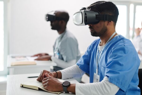
Extended reality (XR) is emerging as a powerful tool in this evolution. Virtual and augmented reality platforms can simulate neuraxial procedures in anatomically accurate environments, allowing learners to:
- Perform repeated placements without patient risk.
- Receive immediate feedback on probe position, needle angle, and target accuracy.
- Experience diverse patient anatomies to build adaptability.
Importantly, these simulations can integrate AI guidance, creating a training environment that mirrors real-world workflows. This fusion of imaging, AI, and simulation allows learners to develop a cohesive procedural mindset — where technology and clinical judgment work in tandem.
Professional societies are adapting to this need. Courses in ultrasound-guided regional anesthesia increasingly emphasize not just basic scanning skills, but the ability to interpret imaging and apply findings in real time — skills essential for modern neuraxial anesthesia.
Continuous education (CE) will enable today’s up-and-coming practitioners to become tomorrow’s medical leaders. And programs such as the Ultrasound-Guided Regional Anesthesia Course offered by ASRA are expanding access to CE:
- Hands-on training equips anesthesiologists with the latest techniques for safer and more efficient procedures.
- Procedural refinement opportunities ensure clinicians can confidently implement new tools and methods.
- Knowledge-sharing platforms facilitate discussions about emerging trends and best practices.
These combined initiatives foster continuous innovation and raise the clinical standard in anesthesiology. By actively pursuing ongoing education and staying current with emerging advancements, clinicians are better equipped to deliver care that reflects the most effective, evidence-based practices available today.
Patient safety and standardization
A persistent challenge in neuraxial anesthesia has been variability — in terminology, technique, and documentation. This creates avoidable risks, especially in collaborative environments where handoffs are common. Clear procedural communication requires standardized language and process definitions.
The ASRA-ESRA Delphi Consensus Study has made significant progress toward terminology harmonization, defining key procedural steps and anatomical descriptors to ensure a common language across institutions and training programs. While implementation varies, the long-term goal is to make standardized language and imaging documentation routine.
Infection control has also come under renewed scrutiny. Updated guidelines emphasize:
- Strict aseptic technique, regardless of setting
- Use of sterile, single-use ultrasound covers
- Tailored protocols for immunocompromised patients or those with prior spinal surgery
When imaging and AI are embedded directly into these protocols, safety is enhanced. Real-time image documentation creates a procedural record that supports quality assurance, legal protection, and training. AI-supported imaging can also detect early deviations from optimal technique, allowing anesthesiologists to avoid potential errors.
Alternative techniques and medications
Ongoing medication shortages — especially for key agents like bupivacaine and opioids—have forced anesthesiologists to rethink their pharmacologic approach to neuraxial anesthesia. These shortages have accelerated the adoption of:
- Multimodal analgesia, reducing reliance on any single agent
- Non-opioid adjuncts like alpha-2 agonists and low-dose ketamine
- Alternative local anesthetics with more reliable supply chains
Professional organizations like the American Society of Anesthesiologists (ASA) have taken proactive roles, issuing real-time shortage tracking and guidance for drug substitution and joining forces with the U.S. Food and Drug Administration (FDA). However, long-term resilience requires a broader rethinking of how medications, techniques, and technology interact.

Technological integration in workflow
Serving patients increasingly falls on individual healthcare practitioners. This dilemma has cast a stark light on anesthesiologists in particular as they grapple with the ramifications of a diminished healthcare infrastructure — especially in rural settings. With fewer skilled anesthetists in the field and less time to allocate per procedure, a fundamental skill now required for anesthesiologist roles will be the ability to do more with less.
The final step toward a smarter future lies in seamless technology integration. To succeed at scale, imaging, AI, and monitoring systems must:
- Integrate directly with electronic health records (EHRs)
- Use single, intuitive interfaces, reducing the learning curve between systems
- Fit into existing ergonomic workflows, preserving operator comfort and efficiency
The goal is for technology to enhance and streamline clinical operations — supporting clinicians without disrupting their cognitive or physical flow. When that happens, imaging, AI, and automation become natural extensions of clinical expertise, rather than technological burdens.
RIVANNA’s Accuro 3S
At RIVANNA, we will continue to push the boundaries of what’s possible for AI-powered ultrasound guidance. In collaboration with Duke University’s Dr. Charles Y. Kim, we have secured a new patent for an advanced ultrasound-guided needle insertion system, reinforcing its commitment to enhancing imaging solutions for spinal needle interventions.
With this patent and ongoing clinical trials, we’ve made continuous strides in research and development for our new AI-powered Accuro 3S platform. This system features a dual-array multi-angle probe that integrates data from two transducer arrays, offering a wide-field, detailed view of the target area. The detachable needle guide ensures consistent alignment within the ultrasound viewing planes, boosting accuracy and reducing the risk of complications.
The split-array design enables an in-plane needle approach through the probe’s center, enhancing accessibility and control. Clinicians benefit from automated needle tracking, allowing for more adaptive and reliable guidance during procedures. Additionally, a hands-free patient drape with supportive bands simplifies workflow, reducing procedural complexity and enhancing ergonomic efficiency.
Follow us for more on the future of neuraxial anesthesia!
Indeed, it’s becoming clear that innovation and adaptability are the framework of anesthesiology’s future. Accuro has stood at the forefront of this evolution, with AI-enabled technology designed to enhance precision and efficiency in anesthesia delivery. By embracing technology, medical professionals affirm their commitment to excellence and to ensuring patient care continually reaches new heights.
The coming year is about more than just keeping pace with change; it’s about leading the charge toward a more promising, reliable, and advanced healthcare landscape.